
Back to Basics – The Retina
Overview of the anatomy of the Retina
The retina is a thin layer of light-sensitive tissue at the back of the eye that covers about 65 percent of its interior surface. It is made up of ten layers as illustrated in the diagram below:
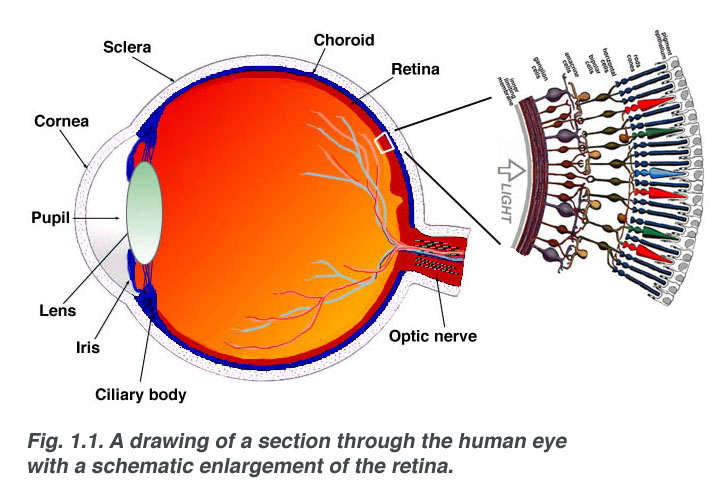
Image from https://lhon.global/eyesight-101/
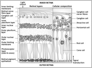
Diagram from http://brain.oxfordjournals.org/content/134/2/518
What does the Retina do?
The purpose of the retina is to receive light that the lens has focused, convert the light into neural signals, and send these signals on to the brain for visual recognition.
The retina is composed of two different kinds of light receptor cells – Rods and Cones. Rods are spread out throughout the retina. There are about 125 million rods in your retina, and these are how you can perceive movement and peripheral vision. In contrast, you have about 6-7 million cones in the very centre of your retina. Very closely concentrated, these provide you with great central vision. Your cones help you see colour and detail.
Do you remember THE dress? The dress that broke the internet because people could not tell if it was blue and black or white and gold? This is actually a really good way of explaining what your rods and cones do: The reason that so many people saw different colours is that the amount of cones in people’s eyes varies. Those with more cones are able to see more clearly and more easily perceive colour differences and details. So if you saw the dress incorrectly as white and gold, or even purple and brown, you have fewer cones than someone who saw blue and black.
The Macular, Optic Nerve & blood vessels of the Retina
Over the next week we will be looking at some disorders of the retina that we see in our clinic at Sydney Ophthalmic Specialists. These often involve the macular or the optic nerve. Here is a brief overview of these structures to help you familiarise with the information we present to you over the coming days.
The Macular is responsible for our central vision & is located in the central retina. The macula is very important as it gives us the vision needed for detailed activities such as reading and writing, and the ability to appreciate colour. The centre of the macular is called the Fovea, it is about 1.5 mm in diameter and is the area where all of the photoreceptors are cones: there are no rods in the fovea. The fovea is the point of sharpest, most acute visual acuity.
Because the fovea has no rods, small dim objects in the dark cannot be seen if one looks directly at them. For instance, to detect faint stars in the sky, one must look just to one side of them so that their light falls on a retinal area, containing numerous rods, outside of the macular zone. Rods detect dim light, as well as movement.
The Optic Nerve is located at the back of the eye & is also known as Cranial Nerve II (2). The job of the optic nerve is to transfer visual information from the retina to the vision centres of the brain via electrical impulses.
The optic nerve is made of ganglionic cells or nerve cells. It consists of over one million nerve fibers. Our blind spot is caused by the absence of specialised light-sensitive cells, or ‘photoreceptors’, in the part of the retina where the optic nerve exits the eye.
Running inferior to the optic nerve is the Central retinal artery. It’s branches spread all over the internal surface of the retina. The central retinal artery supplies the larger part of the retina and all of the nerve fibres that form the optic nerve. Just like any of our other arteries in our body, it too is susceptible to damage & blockages.. 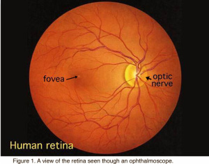
Stay tuned this week as we discuss some of the retinal conditions we treat at Sydney Ophthalmic Specialists.
References
http://hyperphysics.phy-astr.gsu.edu/hbase/vision/retina.html
www.healthline.com/human-body-maps/retina
http://retinahi.com/retina-responsibilties-retina/
http://www.tedmontgomery.com
http://www.healthline.com/human-body-maps/optic-nerve