
Glaucoma – Back to Basics
Glaucoma
The Facts:
- Glaucoma has been described as an ‘invisible disease’.
- It is the leading cause of irreversible blindness worldwide
- 1 in 8 Australians over 80 years of age will develop Glaucoma
- There is a strong hereditary link – first degree relatives of glaucoma patients have a “ten-fold increased risk of developing the disease” (Glaucoma.org)
- 50% of the population who have glaucoma are undiagnosed
What is Glaucoma?
Glaucoma is the name given to a group of eye diseases that cause slow deterioration to the optic nerve at the back of the eye over a period of time. The damage that is done to the optic nerve occurs in most people, as a result of increased pressure inside the eye. Pressure inside the eye, known as intraocular pressure increases when there is a blockage of aqueous circulation or slow drainage of the aqueous. In other patients, damage to the optic nerve may be caused by poor blood supply to the optic nerve fibres. There may also be a weakness in the structure of the nerve and / or a problem in the health of the nerve fibres themselves.
Types of Glaucoma
Primary, open angle glaucoma: This is the most common type of glaucoma. It has very few symptoms until most of the eyesight has been lost at the later stage of the disease. Damage to the vision often develops very slowly with vision gradually fading from the peripheral field in. One eye compensates for the other & the person remains unaware of any problem until many nerve fibres and a significant part of the vision has been destroyed. This damage, caused by glaucoma is irreversible. which is why it is so important to detect the problem as soon as possible and therefor be able to commence treatment as soon as possible with as little damage to the vision as possible. Low pressure or normal pressure glaucoma: Where the intraocular pressure (IOP) may seem to be normal but yet there is damage to the optic nerve Acute, or angle closure glaucoma: Acute angle closure glaucoma is considered an ocular emergency. Symptoms of a painful red eye, blurred vision, headaches, nausea and vomiting alerts the individual to seek medical attention immediately. Congenital glaucoma: Congenital glaucoma is usually diagnosed within the first year of life and is a rare condition. It may be inherited, caused by incorrect development of the eye’s drainage system (the trabecular meshwork) before birth. Symptoms of congenital glaucoma include enlarged eyes, cloudiness of the cornea and sensitivity to light. Both surgery and medication are often required in most cases. Secondary Glaucoma: This can develop as the result of other disorders. Such as injuries, cataracts and eye inflammation.

Examples of different types of glaucoma
Risk factors for glaucoma
While glaucoma is more common as people age, it is important to know that it can occur at any age. Some people are at a higher risk of developing glaucoma than others; these include those people that have:
- a family history of glaucoma
- Diabetes
- Myopia (short sightedness)
- Migraine
- had a previous eye injury
- suffer blood pressure problems
- have a history of prolonged use of cortisone drugs (Steroids)
- and some specific ethnic groups
Diagnosis of Glaucoma
A diagnosis of glaucoma can only be made if a complete eye examination has been performed. Simply checking pressure (IOP) is not enough to conclusively make a diagnosis of glaucoma. A comprehensive eye examination will involve several tests. These include:
- Tonometry: This is the measurement of intraocular pressure. Normal pressure is considered a value between 10-21mmHg.
- Gonioscopy: This is a measurement of your eye’s drainage angle. Using a mirrored lens your ophthalmologist will examine the angle at the front of your eye to determine the type of glaucoma you have; be it open or narrow angle glaucoma.
- Ophthalmoscopy: Your ophthalmologist will use an instrument to examine the back of your eye, paying particular attention to the appearance and health of your optic nerve.
- Visual Field: Tests to examine visual field or peripheral vision looks at the sensitivity of the optic nerve fibres. Any damage to the optic nerve that has occurred as a result of pressure inside the eye will result in a visual field defect.
- Pachymetry: Pachymetry measures the thickness of the cornea. There is a relationship between the thickness of the cornea and the pressure reading. Generally, the thinner the cornea, the higher the pressure reading may be.

A patient has her IOP measured using a lit lamp. Image courtesy of http://www.afb.org
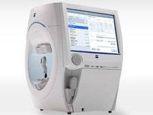
Example of a viusal field analyser – Zeiss
Treatment of Glaucoma
Eye drops are the most common form of treatment for glaucoma. These eye drops work by lowering the pressure inside the eye. Glaucoma medications lower your eye pressure in one of two ways:
- By reducing the amount of fluid created in the eye
- By helping this fluid flow out of the eye through the trabecular meshwork (the drainage angle in the eye).
These eye drops must be taken every day. Just like any other prescription medication, it is important to follow the instructions of your doctor and use your eye drops regularly. In some cases, it necessary to undergo surgery to treat glaucoma. Surgery aims to improve the flow of fluid out of the eye, resulting in lower eye pressure. Glaucoma patients tend to be seen by their ophthalmologist regularly, sometimes as often as every 3-6 months. If you have been diagnosed with glaucoma, have a family history of the disease or any other concerns about your vision, please call us to make an appointment to have an eye examination with one of our ophthalmologists on (02) 9241 2913.
References: